Overview
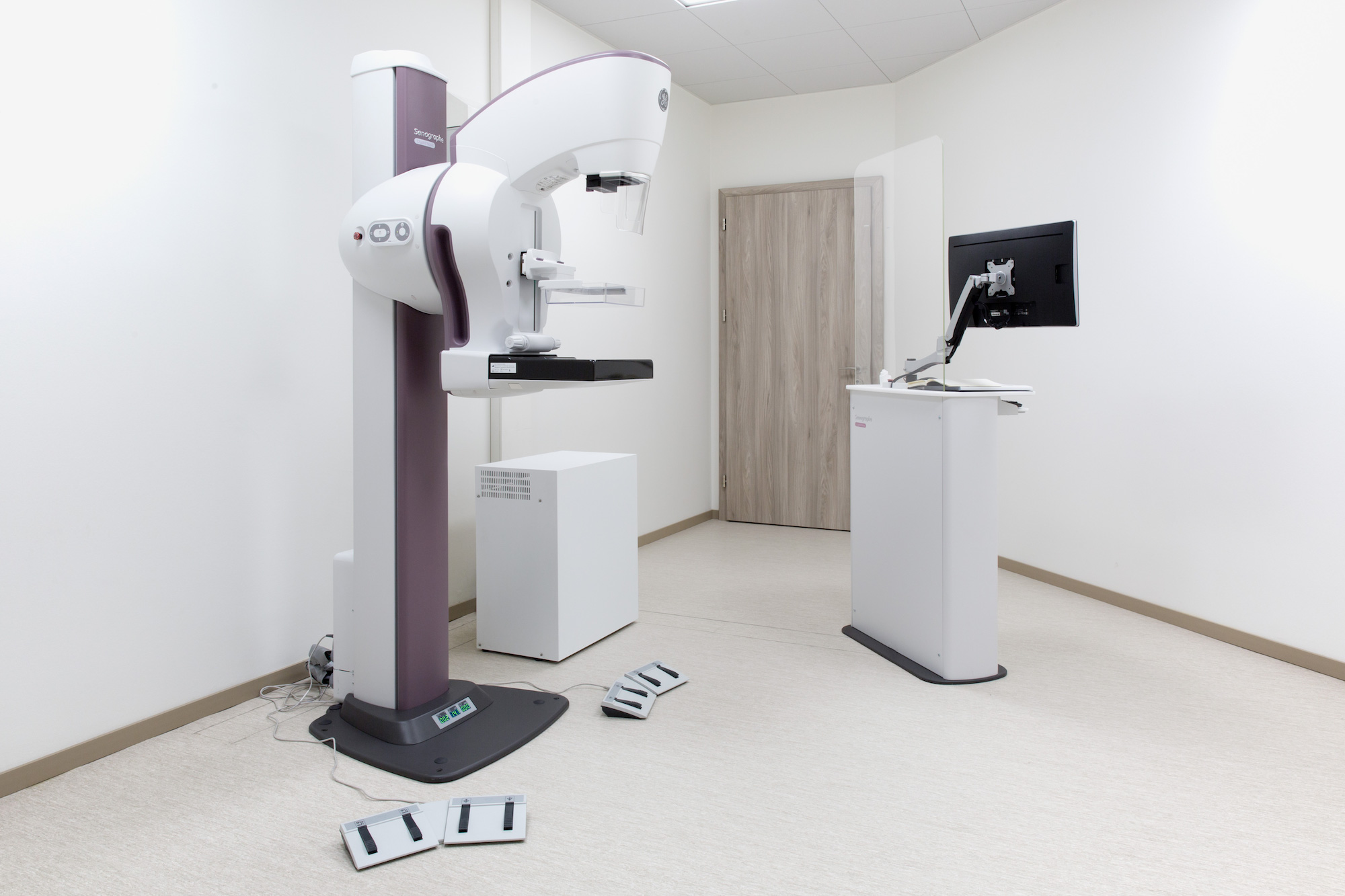
Breast examination is carried out using a special X-Ray device (senographe) which can spot cancers at a very early stage.
The examination process
The breast is placed on the X-ray machine and gently but firmly compressed with a clear plate. The compression only lasts for a few seconds and is necessary to flatten the breast, reducing its density and improving its analysis.
Two X-rays of each breast are taken at different angles.
If necessary, the radiologist might ask for additional X-rays.
If your breasts are dense (rich in glandular tissue), the mammography will have to be completed by a breast ultrasound which will be preformed by the radiologist.
Preparing for a mammography
Your appointment should be scheduled during the first 10 days of your cycle for several reasons :
- The breast is less dense and its analysis is therefore easier
- Breast compression is better tolerated
- You are certainly not pregnant
If you had previous mammographs, please bring them with you.

